

For centuries, the study of human anatomy was confined to the dissection of cadavers and the study of two-dimensional textbook illustrations. While these methods laid the foundation for modern medicine, they presented significant limitations in accessibility, repeatability, and the visualization of complex spatial relationships within the human body.
Today, the landscape of medical education, surgical planning, and patient care is undergoing a massive transformation, driven by the rapid advancement of 3D anatomical models. Whether in the form of highly interactive digital software or precisely 3D-printed physical replicas, these models are bridging the gap between theoretical knowledge and practical application.
Here is an in-depth look at how 3D anatomical models are reshaping the healthcare and educational sectors, the different technologies driving them, and what the future holds for this incredible innovation.
The Evolution of Anatomical Study
To understand the value of 3D anatomical models, we must look at the traditional challenges of learning anatomy.
Historically, cadaveric dissection has been the gold standard for medical students. However, cadavers are expensive to maintain, limited in supply, and only offer a single-use learning experience. Once a structure is dissected, it cannot be undone for another student to practice on. Furthermore, 2D textbook images, while detailed, force the brain to do the heavy lifting of translating a flat image into a complex 3D mental construct.
The introduction of physical plastic models helped, but they were often generalized, lacking the intricate details and physiological variations present in real human patients. The advent of modern 3D modeling—both digital and physical—has solved these problems by offering infinite repeatability, flawless accuracy, and unprecedented customization.
The Two Pillars of 3D Anatomical Models
When we talk about 3D anatomical models today, we are generally referring to two distinct but highly complementary technologies: Virtual/Digital Models and Physical 3D-Printed Models.
1. Digital and Virtual 3D Models (Software, VR, and AR)
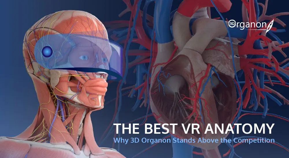
Digital models are highly detailed, computer-generated representations of the human body. Accessible via tablets, computers, or virtual reality (VR) headsets, these platforms allow users to explore the body layer by layer.
- Interactive Learning: Medical students can use software to strip away skin, muscle, and bone to view the cardiovascular or nervous systems in isolation. They can rotate the heart 360 degrees, zoom in on the valves, and even watch animated simulations of blood flow.
- Virtual Reality (VR) and Augmented Reality (AR): VR takes digital models a step further by immersing the user inside the anatomy. Surgeons can literally “walk through” a virtual model of a patient’s brain to map out a tumor extraction. AR allows instructors to project a beating 3D heart onto a desk in front of a classroom, combining the digital model with the physical environment.
2. Physical 3D-Printed Models
While virtual models are excellent for visual learning, physical 3D-printed models provide the essential element of tactile feedback (haptics). Using data from a patient’s CT scan or MRI, medical professionals can now print a 1:1 scale, exact physical replica of that specific patient’s organs or bones.
- Multi-Material Printing: Advanced 3D printers do not just print hard plastics. They can use multiple materials simultaneously to mimic the distinct textures of human tissue. A single printed model of a kidney can feature a hard plastic tumor, surrounded by soft, rubbery tissue, complete with flexible, color-coded blood vessels.
Key Applications Transforming Healthcare
The integration of 3D anatomical models extends far beyond the university lecture hall. They are actively saving lives and improving outcomes in clinical settings every single day.
Surgical Planning and Simulation
Perhaps the most profound impact of 3D models is in preoperative planning. In the past, surgeons had to rely on 2D scans to plan complex procedures, often encountering unexpected anatomical variations only after the patient was opened up on the operating table.
Today, a surgeon can request a 3D-printed model of a patient’s exact spine, heart, or skull. They can physically hold the patient’s anatomy in their hands, evaluate the problem from every angle, and even perform a “practice run” of the surgery on the model using actual surgical tools. This level of preparation reduces time spent in the operating room, minimizes blood loss, lowers the risk of complications, and leads to significantly better patient outcomes.
Medical Device Testing and Customization
Before a new pacemaker, stent, or joint replacement is approved for human use, it must be rigorously tested. 3D anatomical models provide a safe, accurate testing ground for these devices. Furthermore, rather than relying on mass-produced, one-size-fits-all implants, doctors can use 3D models to design and manufacture custom-fit prosthetics and implants tailored perfectly to the patient’s unique anatomy.
Enhancing Patient Communication
Medical jargon and black-and-white MRI scans can be incredibly intimidating and confusing for patients. When a doctor needs to explain a complex condition—such as a congenital heart defect or a complicated bone fracture—handing the patient a physical 3D model of their own organ bridges the communication gap. It allows the patient to clearly see the problem and understand the proposed surgical solution, leading to higher patient confidence and informed consent.
The Future of 3D Anatomical Modeling
The technology surrounding 3D anatomy is advancing at a breakneck pace. As we look to the near future, several exciting trends are emerging:
- Artificial Intelligence (AI) Integration: AI algorithms are dramatically reducing the time it takes to convert 2D medical scans into 3D printable files. What used to take hours of manual digital segmentation can now be done by AI in minutes.
- Bioprinting: The ultimate frontier of 3D anatomical modeling is bioprinting—using “bio-ink” made of living human cells to print functional, living tissue. While printing fully functional, transplantable organs is still years away, scientists are already successfully printing patches of tissue for drug testing and research.
- Widespread Accessibility: As 3D printers and VR headsets become more affordable, we will see these tools move from elite research hospitals into everyday clinical practices and high school biology classrooms.
Conclusion
3D anatomical models are not just a technological novelty; they represent a fundamental paradigm shift in how we understand, interact with, and heal the human body. By providing unparalleled accuracy, patient-specific customization, and risk-free environments for surgical simulation, both digital and physical 3D models are setting a new, elevated standard of care. For medical professionals, educators, and patients alike, the ability to hold, rotate, and interact with the complexities of human anatomy in three dimensions is truly a medical marvel.